
Adenium Flower Streaks: What Virus Evidence Can Tell You
Adenium flower streaks can reflect virus evidence, but a bloom pattern is not a diagnosis. Compare tissue, contain spread, and test carefully.
Maya Ellison · 2026-09-03 · Updated 2026-10-24 · 13 min read
Key Takeaways
- The 2025 PFBV report supports a virus-associated flower-break signal in Adenium, including graft transmission, but it does not make every streaked bloom a confirmed infection.
- HoCSV is an earlier experimental example in which flower color break followed systemic infection, so the symptom deserves context rather than instant certainty.
- Compare newly opened blooms and new leaves with a same-plant reference, then record dates, images, recent changes, and plant source before drawing a conclusion.
- Pause propagation and use a clean-tool gate when sap or residue could move between plants. Quarantine and laboratory testing are more useful than a symptom-only label.
- A negative or incomplete home observation cannot rule out infection. Sampling quality, assay scope, and the timing of tissue collection all matter.
Streaks in an Adenium flower can be a virus clue, but they cannot name the virus.
A 2025 report confirmed Pelargonium flower break virus in Adenium obesum plants from Brazil. Earlier work had already shown that Hoya chlorotic spot virus can infect experimental desert rose and produce a later flower color break.
The practical response is to document the pattern, pause propagation, reduce tool contact, and seek a target-appropriate diagnosis.
The new finding expands the list of plausible causes. It does not show that every streaked desert rose is infected or that PFBV is common in US collections.
A petal photograph cannot distinguish PFBV from HoCSV, cucumber mosaic virus, cultivar variation, or environmental injury.
What the New PFBV Report Changes
What the researchers found
Researchers observed Adenium obesum plants with virus-like symptoms at two locations in Brazil. High-throughput RNA sequencing from one sample produced a 3,923-nucleotide contig with 97.77% nucleotide identity to Pelargonium flower break virus. Real-time RT-PCR then confirmed PFBV in both samples.
The researchers also tested transmission routes. Sap inoculation to healthy Adenium seedlings and Pelargonium zonale did not succeed in the reported attempts. Grafting transmitted PFBV from and to an Adenium plant.
The 2025 paper established a natural host record supported by molecular detection, not a prevalence estimate based on flower photographs.
What the report does not establish
A first natural-host report answers one narrow question. It shows that the virus was detected in a naturally infected host under the conditions described by the researchers.
It does not measure how often the virus occurs in Adenium collections.
It also does not establish a US distribution, a general crop-loss estimate, or a universal symptom pattern. The transmission result is pathway-specific. Successful grafting does not prove that every pruning contact transmits PFBV, and failed sap inoculation does not prove that all mechanical contact is harmless.
Use the paper as a reason to investigate a suspicious bloom. Do not use it as permission to label every pale stripe PFBV.
Why HoCSV Belongs in the Same Conversation
The earlier experimental flower pattern
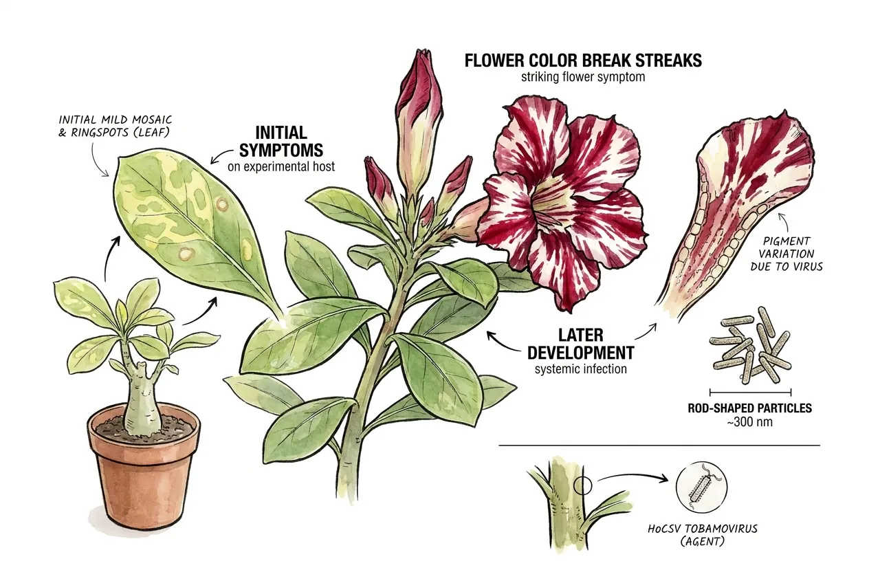
The HoCSV paper began with virus-like symptoms on Hoya. Researchers mechanically transmitted the agent, observed rod-shaped particles about 300 nanometers long, and characterized a novel tobamovirus with a 6,386-nucleotide genome.
In its experimental host range, desert rose developed systemic infection. The paper describes initial mild mosaic and ringspots on leaves that faded over time, followed later by a striking flower color break.
This sequence explains why a bloom can be part of a plant-wide virus investigation even when the earlier leaf pattern is no longer obvious.
HoCSV makes a streaked Adenium flower biologically plausible as part of a virus symptom sequence, but it still does not identify the agent in a new plant.
Why the two reports are not interchangeable
The studies used different evidence paths. One identified PFBV in naturally infected Adenium samples and reported graft transmission. The other experimentally infected plants with HoCSV and observed a later flower symptom in the host-range work.
| Virus record | Main evidence in the report | What it supports | What it does not prove |
|---|---|---|---|
| PFBV | Sequence match, RT-qPCR, and graft transmission | Adenium can be a natural host | That every streak is PFBV |
| HoCSV | Experimental infection, genome work, and host-range symptoms | HoCSV can infect tested desert rose | That a home plant carries HoCSV |
| CMV | Serology plus biological and structural confirmation | CMV has an Adenium record | That a flower streak is CMV |
The table is a map of evidence types. It is not a symptom key. Virus identity still depends on a suitable sample and assay.
How to Read a Streaked Adenium Flower
A streak is a signal, not a test
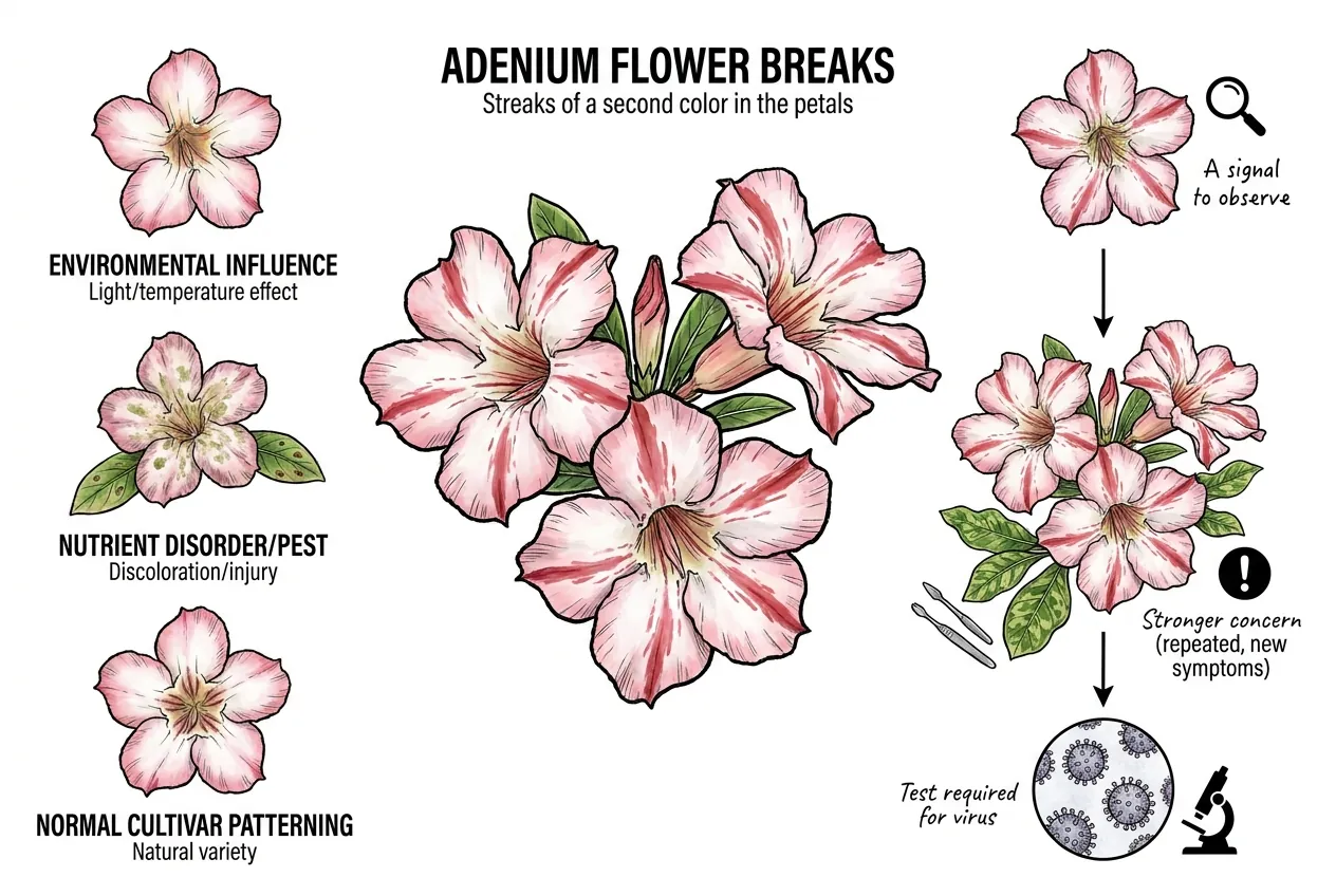
Plant-virus guidance describes flower breaks as streaks of a second color in the petals. The same guidance says symptoms vary with the virus and the plant. That matters for Adenium because cultivar, tissue age, environmental conditions, and mixed infections can change what a grower sees.
Other plant problems can also change color or form. Light and temperature can influence symptom expression. Nutrient disorders, pests, chemical injury, and normal cultivar patterning can overlap with a virus-like appearance.
Treat a single unusual bloom as a signal to observe. Stronger concern comes from repeated changes on new blooms, new leaf mosaic or ringspots, or a history of shared propagation work. None of those observations alone names PFBV or HoCSV.
Simple check before the bloom fades
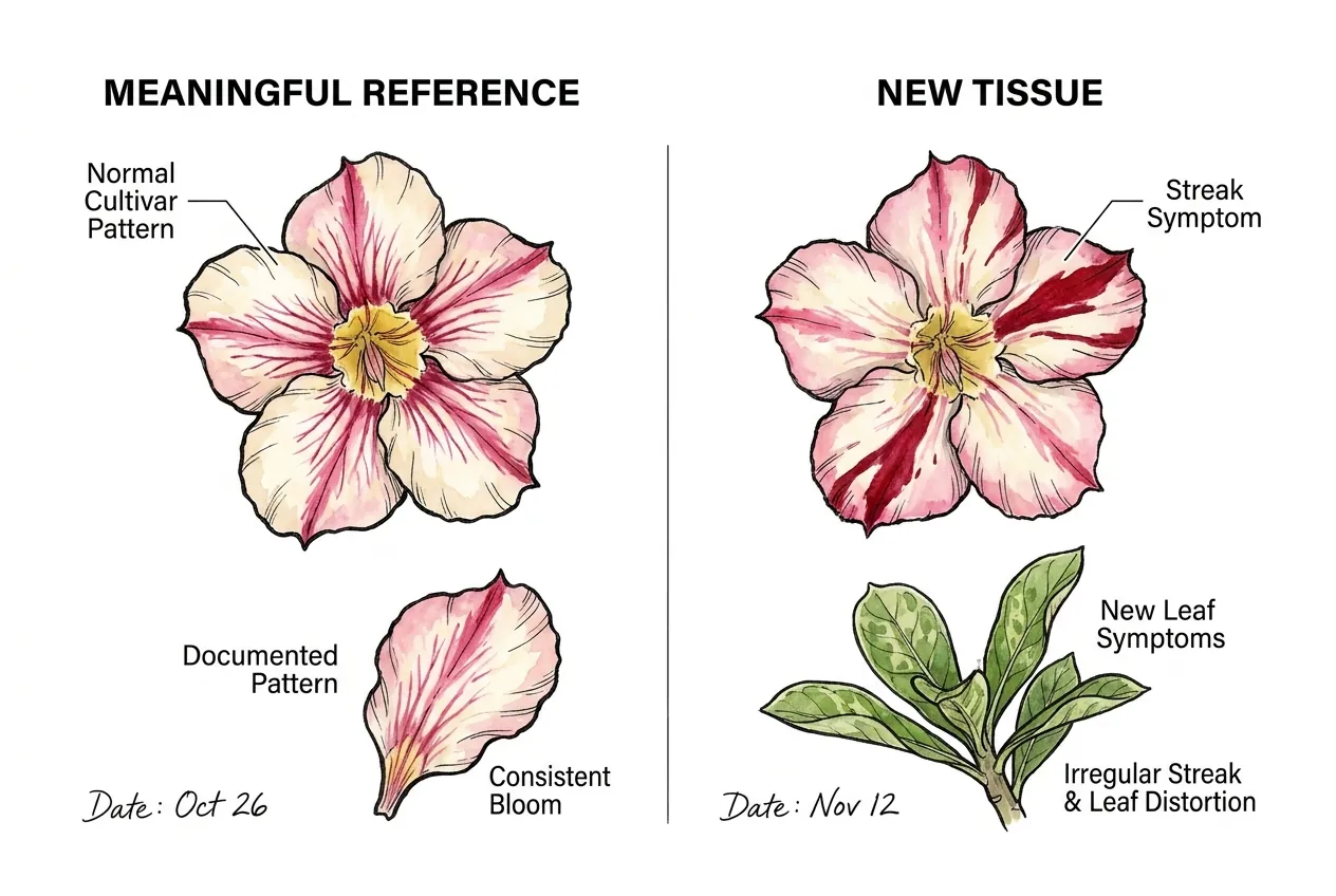
Give the plant a unique tag and photograph the whole plant, the newly opened bloom, and the newest leaves before cutting or changing its treatment. Compare the bloom with an earlier photograph of the same plant at the same opening stage. An unrelated cultivar is a poor color reference.
For the next three blooms, photograph within 24 hours of full opening, using the same background, indirect daylight, and camera settings. Put a white card and ruler beside the flower, without touching it. Three blooms are a manageable observation set that helps separate a single damaged flower from a recurring pattern. They cannot clear the plant of infection.
Record each bloom’s opening date and any new leaf mosaic or ringspots. Do not wait for three blooms if leaf symptoms appear, shared grafting is planned, or several plants develop a similar change. Keep the plant out of propagation work and contact a diagnostic clinic.
Compare blooms from the same branch
On a grafted plant, identify the branch that produced each bloom. Photograph the branch back to its graft junction without cutting it, and put that branch identifier in the image filename. Flowers from different scions are not a valid before-and-after color comparison.
Use a pot label or a loose tag on the support rather than a tight tie around a swelling shoot. If the branch history is unknown, start a new baseline for that branch. This keeps cultivar differences from becoming a false symptom trend while preserving a useful record for the clinic.
What Other Adenium Virus Records Add
CMV explains why leaf symptoms need their own evidence
The USDA record for cucumber mosaic virus described desert rose leaves with mosaic, dark-green islands around veins, and chlorosis at the margins. CMV was confirmed by serological testing.
The investigators also mechanically transmitted an agent to Chenopodium quinoa, observed chlorotic local lesions, and used dsRNA analysis and electron microscopy. The reported particles were about 28 nanometers across.
A separate Taiwan report described mosaic, chlorotic ringspots, and line patterns in Adenium. These reports support CMV as a documented Adenium virus association. They do not make CMV the default explanation for flower streaks.
The potexvirus record widens the differential
A 2023 study described desert rose mottle virus from symptomatic Adenium collected in several regions of Brazil.
Researchers reproduced mottle symptoms in mechanically inoculated A. obesum and determined a 6,781-nucleotide genome through high-throughput sequencing and Sanger confirmation.
A separate 2023 report identified Adenium obesum virus X in A. obesum submitted to Australian post-entry quarantine after import from China.
That genome record widens the named differential, but it does not establish that AobVX causes flower color breaks.
The practical lesson is narrow but important. A CMV-specific negative test does not rule out every virus, and a plant with persistent mottle may need a broader diagnostic question.
The study did not establish that this potexvirus causes the flower color break described here.
Record leaf mosaic and chlorotic rings separately from flower streaks. Adenium Cucumber Mosaic Virus Field ID and Triage explains the CMV-specific checks.
What to Do Before Testing
Pause propagation and preserve context
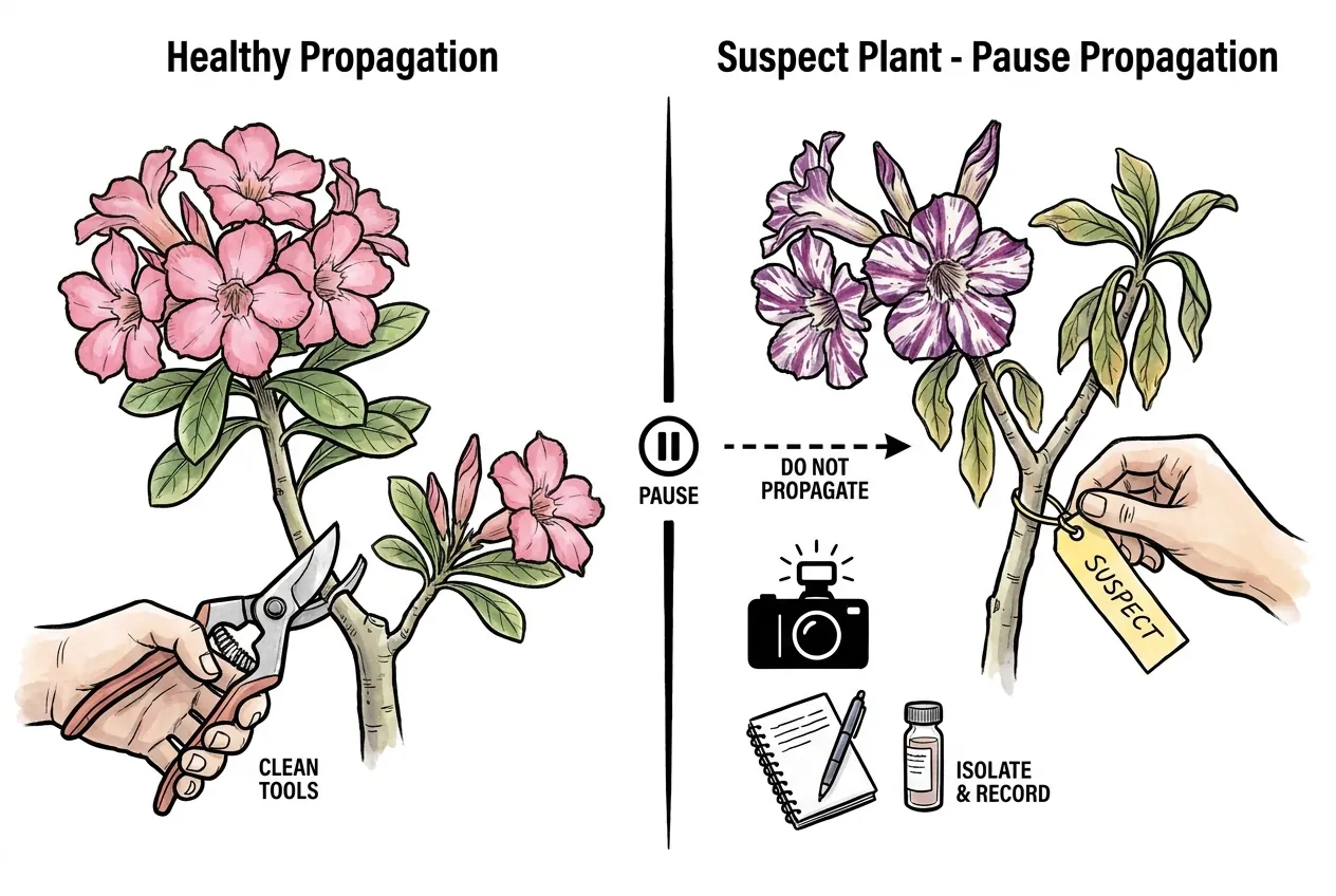
Do not use a suspect plant as a scion, rootstock, or cutting source while the question matters.
Label it and keep it apart from routine propagation work. Photograph the bloom before it fades and record recent handling.
If a necessary cut cannot wait, handle the suspect plant separately from healthy plants. Keep sap and debris away from shared work surfaces.
Isolation does not prove infection, and it does not cure an infected plant. It simply reduces avoidable exposure while the evidence is incomplete.
Handle Adenium sap carefully
Adenium sap is poisonous and can irritate skin. Wear suitable hand and eye protection when a cut or sap contact is unavoidable. Keep sap and plant pieces away from children and pets, avoid touching your eyes or mouth, and wash exposed skin with soap and water.
Use a clean-tool gate
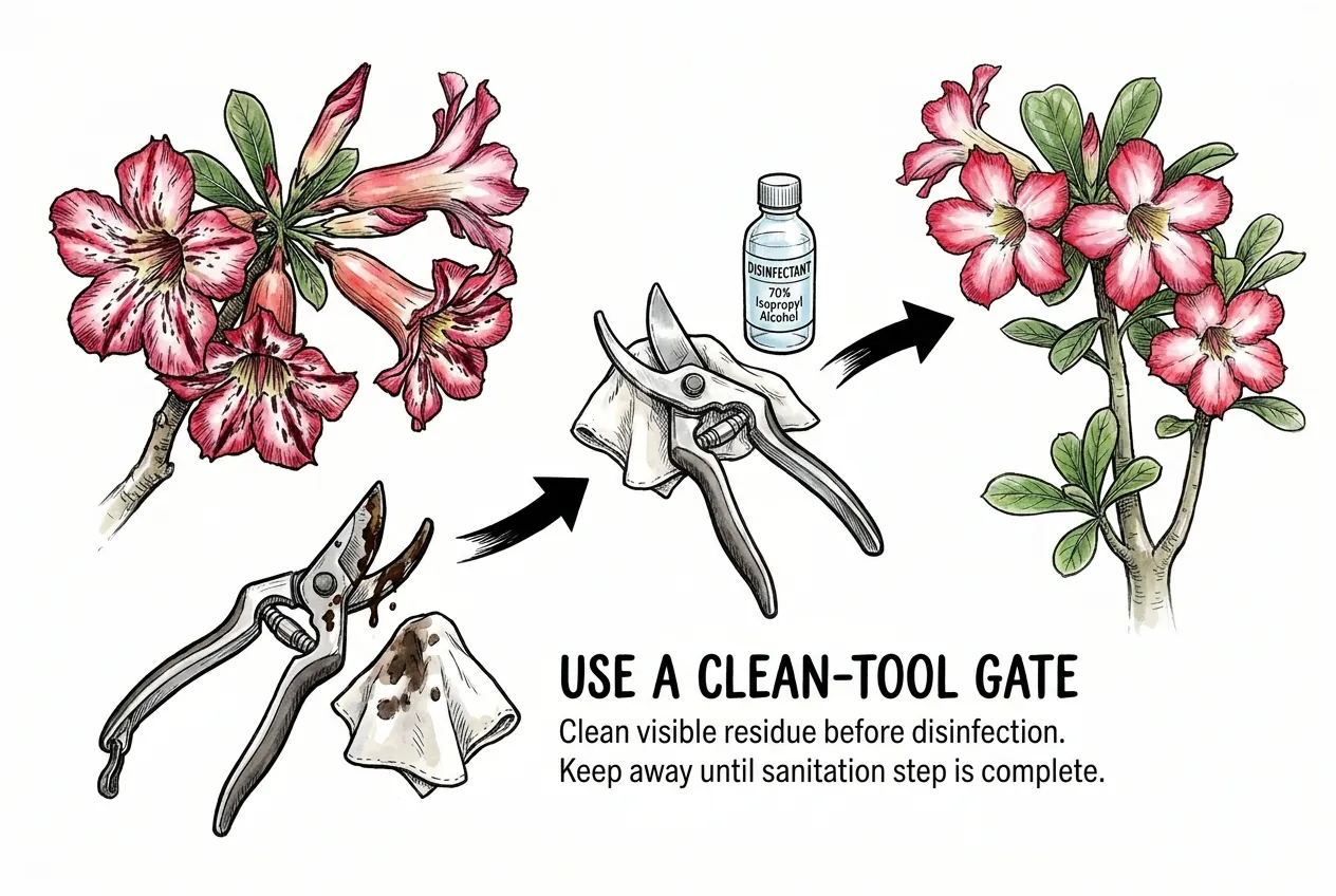
Keep a dedicated cutting tool with the suspect plant. If a cut is essential, work on that plant last, contain sap and debris, and keep the tool out of the clean-tool area afterward. Clean visible residue before disinfection.
Do not treat a quick alcohol wipe as verified control for every possible virus. Ask the diagnostic laboratory for an appropriate sanitation method and follow the selected product’s labeled dilution, wet contact time, and rinse instructions. If its suitability is unresolved, keep the dedicated tool separate instead of reusing it.
Use two labeled containers for clean and used tools. A tool returns to the clean container only after cleaning and the complete disinfection procedure. This physical separation prevents a cleaned-looking blade from accidentally moving to the next plant.
What a Real Diagnosis Looks Like
Measured check with a sample and contact record
Create one row per plant with its tag, first symptom date, bloom count, leaf findings, last shared-tool session, and any graft partners. Include contacts since the last documented cleaning rather than guessing a virus incubation period. Handle healthy plants first and unresolved plants last.
Before cutting a sample, send the clinic the photographs and ask whether its service tests PFBV and HoCSV, or only common targets such as CMV. Record the assay name, requested tissue, amount, packaging, and submission date on the same row. Follow the clinic’s sampling instructions because the correct tissue and quantity depend on the assay.
For example, a report saying CMV not detected belongs in the CMV result field, while PFBV and HoCSV remain untested. If the laboratory lacks those assays, retain the isolation and propagation pause while discussing another laboratory or broader testing. Do not turn a limited negative into virus-free status.
Read a negative result narrowly
An RT-PCR or immunostrip result answers a question about its target, sample, and method. A negative CMV screen can reduce concern about CMV when the test and sample were suitable.
It cannot rule out PFBV, HoCSV, a potexvirus, another pathogen, or an abiotic cause.
The PFBV report used sequencing and real-time RT-PCR. The HoCSV paper developed specific coat-protein primers. The CMV record used serology and additional biological and structural evidence.
Match every result to the plant tag, sampled tissue, collection date, and exact test target. A result from a different plant or an old sample cannot clear today’s symptomatic growth.
When symptoms persist after a negative single-target result, keep the plant separate and ask whether a broader assay or a different sample is appropriate.
Do not turn one negative result into the statement that the plant is virus-free.
A negative result is evidence about one tested target under one method, not a universal clearance certificate.
A Calm Decision Path for a Suspect Bloom
The first useful record is the bloom’s history. Photograph the whole plant and new tissue under consistent conditions, then note whether the pattern is normal for the same plant or cultivar.
If the pattern repeats, appears with new leaf symptoms, or matters because the plant will be propagated, isolate it from propagation work. Use a clean-tool gate for any necessary handling. Then ask a diagnostic clinic which tissue and assay fit the case.
If testing identifies a virus, keep the conclusion specific to that virus and follow the laboratory or plant-health authority’s management advice.
If testing is negative for one target but symptoms continue, keep the differential open.
Flower streaks justify better evidence and safer handling. They do not justify certainty from appearance alone.